Retrobulbar haematoma / orbital compartment syndrome
sight-saving canthotomy · Trauma
Physician-written · Verified by three sets of doctors · Reviewed November 2026
Bedside summary
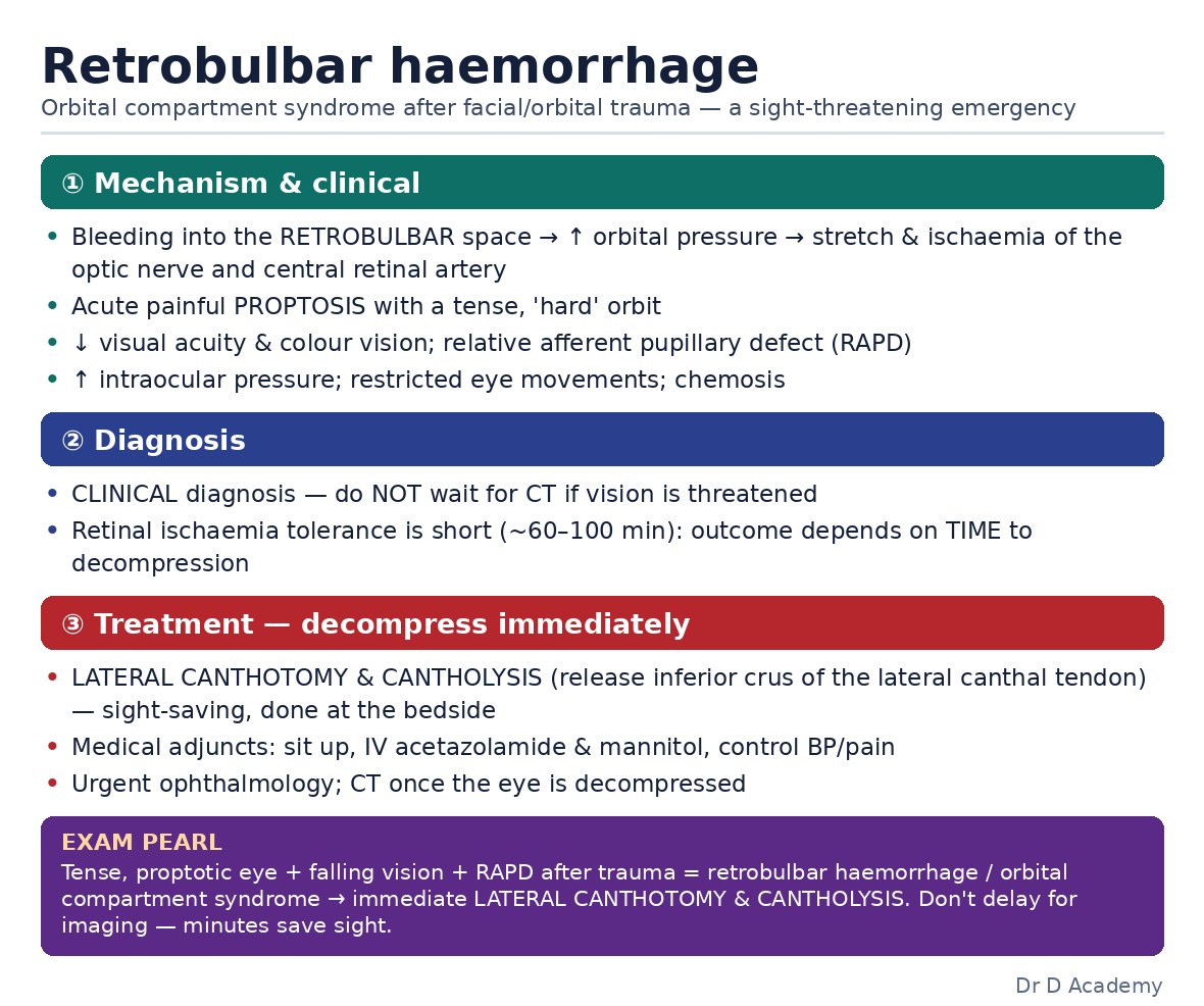
Haemorrhage into the closed orbital space raises intraorbital pressure, stretching the optic nerve and occluding its blood supply. Vision loss is reversible only with emergency lateral canthotomy and cantholysis within minutes to hours.
Figures & algorithms
Red flags
- Relative afferent pupillary defect after facial trauma
- Rapidly falling visual acuity
- Tense proptosed globe that resists retropulsion
- Loss of retinal arterial pulsation/cherry-red macula
- Increasing pain and ophthalmoplegia
Assessment
Differential diagnosis
- Orbital fracture with haemorrhage
- Retrobulbar bleed post-op/procedure
- Globe rupture
- Traumatic optic neuropathy
- Orbital cellulitis/abscess
- Carotid-cavernous fistula
Investigations
- Visual acuity and relative afferent pupillary defect
- Intraocular/orbital pressure (tense, proptosed globe)
- Fundoscopy (disc pallor, central retinal artery pulsation)
- CT orbits once sight-threatening emergency excluded/treated
- Assess extraocular movements
- Colour vision if cooperative
Management
- Recognise the triad: acute proptosis, decreased vision, and a tense, painful orbit after trauma/surgery
- Check for a relative afferent pupillary defect and loss of visual acuity (markers of optic nerve compromise)
- Do not delay for CT if clinical orbital compartment syndrome with vision threat is evident
- Perform immediate lateral canthotomy: inject local anaesthetic, clamp then incise the lateral canthus horizontally
- Perform inferior cantholysis: strum and release the inferior crus of the lateral canthal tendon to decompress
- Reassess intraorbital tension, proptosis, vision, and pupil response after decompression
- Perform superior cantholysis if pressure remains elevated
- Give adjuncts to lower intraorbital/intraocular pressure (elevate head, acetazolamide, mannitol) as a bridge
- Obtain urgent ophthalmology and maxillofacial review
- Image with CT once the eye is decompressed to define the fracture/haematoma
- Treat associated injuries and monitor for re-accumulation
Drugs & doses
Lidocaine with adrenaline local infiltration for canthotomy
Acetazolamide 500 mg IV
Mannitol 1-2 g/kg IV
Dexamethasone (adjunct per ophthalmology)
Disposition
Emergency ophthalmology and maxillofacial surgery; theatre if ongoing haemorrhage; admit and monitor vision and for re-accumulation.
Complications & pitfalls
- Permanent blindness from optic nerve ischaemia
- Central retinal artery occlusion
- Globe injury during the procedure
- Recurrent haematoma
- Incomplete decompression
Exam pearls
- This is a clinical diagnosis: do not wait for imaging when vision is threatened
- The retinal circulation tolerates only ~60-90 minutes of ischaemia, act within minutes
- Cantholysis (releasing the tendon crus), not just the skin cut, achieves decompression
- A relative afferent pupillary defect plus a tense proptotic orbit is the key decision trigger
Disclaimer: Educational decision support for qualified clinicians and students — not a substitute for clinical judgement, local protocols or a full drug reference. Confirm every dose and contraindication against your formulary before prescribing.