Scaphoid fracture (including suspected/occult scaphoid fracture)
scaphoid carpal snuffbox FOOSH avascular-necrosis · Musculoskeletal
Physician-written · Verified by three sets of doctors · Reviewed November 2026
Bedside summary
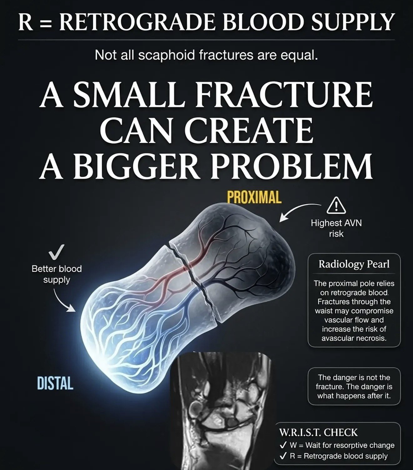
Commonest carpal fracture, typically after a FOOSH in young adults. The scaphoid's retrograde blood supply means proximal/waist fractures risk avascular necrosis and non-union. Initial X-rays miss up to 20-30% of fractures, so clinically suspected fractures (anatomical snuffbox tenderness) are immobilised and re-imaged even with normal radiographs. A missed scaphoid fracture is a classic medicolegal pitfall.
Figures & algorithms
Red flags
- Displaced or proximal pole fracture (AVN / non-union risk)
- Persistent snuffbox tenderness despite normal X-ray (treat as fracture)
- Perilunate/lunate dislocation on lateral view
- Median nerve symptoms or significant swelling
- Delayed presentation with established non-union
Assessment
Differential diagnosis
- Distal radius fracture
- Other carpal fracture (triquetrum, lunate) or perilunate dislocation
- Scapholunate ligament injury
- De Quervain tenosynovitis / wrist sprain
- First CMC joint injury
Investigations
- Clinical signs: anatomical snuffbox tenderness, tenderness over scaphoid tubercle, pain on axial loading of thumb, reduced grip
- Scaphoid-series X-rays (PA, lateral, oblique, PA in ulnar deviation)
- If X-ray normal but clinically suspected: immobilise and either MRI (most sensitive) within days or repeat X-ray at 10-14 days [verify local pathway]
- CT as alternative where MRI unavailable / to assess displacement and union
Management
- Assess for snuffbox and scaphoid tubercle tenderness and thumb axial-load pain.
- Confirmed undisplaced fracture: below-elbow cast (thumb inclusion varies by local protocol), orthopaedic follow-up.
- Displaced fracture (>1-2 mm), proximal pole, or associated instability: refer for surgical fixation (screw) - higher non-union/AVN risk.
- Suspected/occult fracture with normal X-ray: immobilise in a cast/splint and arrange definitive imaging (MRI preferred) or re-X-ray at ~2 weeks; do not simply discharge as a 'sprain'.
- Analgesia and elevation.
- Document and communicate the follow-up plan clearly to the patient (missed scaphoid risk).
- Advise on cast care and return if increasing pain/neurovascular symptoms.
Drugs & doses
Paracetamol 1 g PO every 6 h (max 4 g/day)
Ibuprofen 400 mg PO every 8 h with food (if no contraindication)
Codeine 30-60 mg PO every 6 h for breakthrough pain [verify local dose]
Disposition
Discharge in cast/splint with scaphoid/fracture-clinic follow-up and planned definitive imaging. Refer displaced/proximal fractures for surgical consideration.
Complications & pitfalls
- Non-union
- Avascular necrosis of the proximal pole
- Scaphoid non-union advanced collapse (SNAC wrist) and osteoarthritis
- Chronic wrist pain and reduced function
Exam pearls
- Normal initial X-ray does NOT exclude a scaphoid fracture - immobilise and re-image if clinically suspected.
- MRI is the most sensitive follow-up test and allows early return to activity if negative.
- Proximal pole and displaced fractures have the worst blood supply - low threshold for surgery.
- Document the safety-net plan; missed scaphoid fracture is a common complaint/claim.
- Thumb inclusion in the cast is debated - follow local orthopaedic preference.
Disclaimer: Educational decision support for qualified clinicians and students — not a substitute for clinical judgement, local protocols or a full drug reference. Confirm every dose and contraindication against your formulary before prescribing.