Retinal detachment
Sight-threatening · Ophthalmology
Physician-written · Verified by three sets of doctors · Reviewed November 2026
Bedside summary
Separation of the neurosensory retina — flashes, floaters and a progressing visual-field “curtain”. Macula-on detachment is time-critical for surgery.
Figures & algorithms
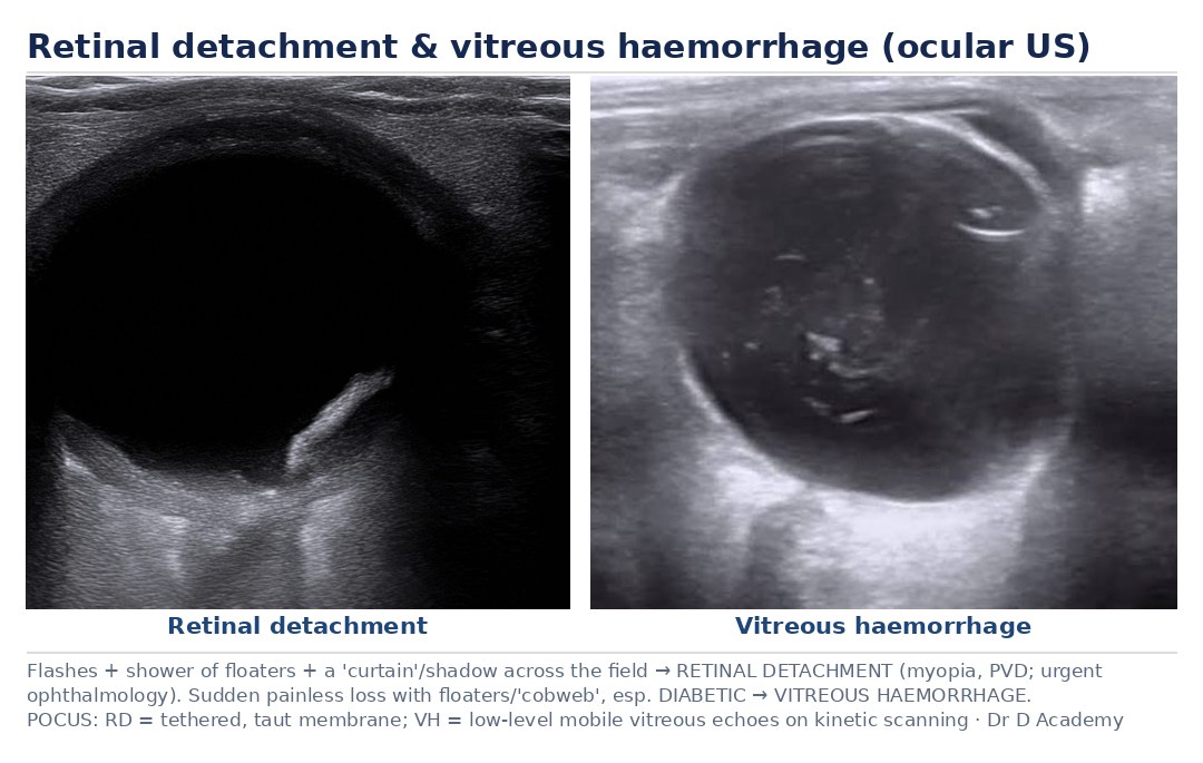
Red flags
- Sudden shower of floaters + flashes + curtain/shadow
- Central vision loss (macula involved)
Assessment
Differential diagnosis
- Posterior vitreous detachment
- Vitreous haemorrhage
- CRVO
- Migraine aura
Investigations
- Visual acuity and fields
- Fundoscopy/slit lamp (detached retina)
- Urgent ophthalmology / B-scan ultrasound
Management
- Urgent ophthalmology referral (same day)
- Macula-on (central vision intact) is more time-critical — expedite
- Advise to keep still; position per ophthalmology advice
- Surgical repair (vitrectomy/buckle/pneumatic retinopexy)
Disposition
Urgent ophthalmology/vitreoretinal team.
Complications & pitfalls
- Permanent vision loss if macula detaches
- Proliferative vitreoretinopathy
Exam pearls
- Flashes + floaters + a curtain = detachment until proven otherwise
- “Macula-on” cases need the most urgent surgery to save central vision
Same-day ophthalmology; expedite macula-on cases for surgery.
Disclaimer: Educational decision support for qualified clinicians and students — not a substitute for clinical judgement, local protocols or a full drug reference. Confirm every dose and contraindication against your formulary before prescribing.