Hyphaema (traumatic)
Trauma · Ophthalmology
Physician-written · Verified by three sets of doctors · Reviewed November 2026
Bedside summary
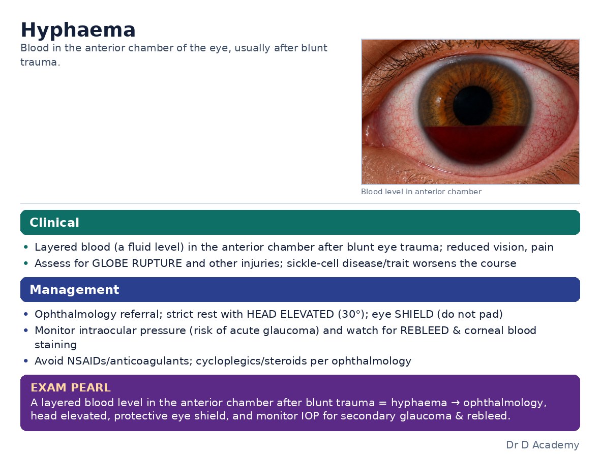
Blood in the anterior chamber after blunt trauma. Risk of raised IOP and rebleeding (esp. days 3–5); consider sickle-cell status.
Figures & algorithms
Red flags
- Raised IOP
- Large hyphaema / total (“8-ball”)
- Sickle-cell disease/trait
- Signs of globe rupture
Assessment
Differential diagnosis
- Globe rupture
- Traumatic iritis
- Vitreous haemorrhage
Investigations
- Visual acuity; measure IOP; slit lamp (blood level)
- Exclude globe rupture; CT if needed
- Sickle screen (affects management)
Management
- Exclude globe rupture first
- Eye rest, head elevation ~30°, protective shield
- Control IOP; avoid NSAIDs/antiplatelets (rebleed risk)
- Urgent ophthalmology; admit/monitor large hyphaema or sickle patients
- Cycloplegic/steroid drops per ophthalmology
Drugs & doses
IOP-lowering agents as needed
Cycloplegic/steroid drops (ophthalmology-directed)
Disposition
Ophthalmology; admit large hyphaema/sickle.
Complications & pitfalls
- Rebleeding, raised IOP/glaucoma
- Corneal blood staining
- Vision loss
Exam pearls
- Always exclude globe rupture
- Head up + shield + rest; avoid antiplatelets/NSAIDs
- Sickle patients tolerate raised IOP poorly
Exclude rupture, rest head-up with a shield, control IOP, avoid NSAIDs, and refer to ophthalmology.
Disclaimer: Educational decision support for qualified clinicians and students — not a substitute for clinical judgement, local protocols or a full drug reference. Confirm every dose and contraindication against your formulary before prescribing.